Arthrodesis, a surgical procedure that involves the permanent fusion of a joint, is performed on dogs to treat severe joint issues. The goal of arthrodesis is to restore partial function of the limb by stabilizing the joint and eliminating pain caused by the joint’s movement. This operation is generally considered a last resort when all other treatment options, such as medication, physical therapy, or less invasive surgeries, have failed to provide relief or restore functionality. At clinics like Vets In The City, this procedure is carefully considered and performed by expert veterinarians to improve the quality of life for affected pets.
Joint arthrodesis is typically recommended for dogs suffering from severe joint damage, chronic pain, or conditions that cannot be treated through standard surgical repair or rehabilitation. This procedure is especially useful for older dogs or dogs with advanced joint disease, as it allows them to regain some level of mobility while eliminating the discomfort caused by the affected joint.
What is Arthrodesis?
Arthrodesis involves the surgical removal of cartilage from a joint, followed by the fusion of the bones to prevent any future movement in that joint. The procedure may seem extreme, but it often provides significant relief for dogs that would otherwise experience ongoing pain and limited use of the limb. The fusion of the bones stops further deterioration of the joint and allows the dog to move in a more stable and pain-free manner, despite the joint’s immobilization.
It is important to note that once a joint is fused, the dog will lose the natural range of motion in that area. However, in most cases, the animal compensates for the loss of mobility and can function normally in its daily life with some adjustments.
Types of Arthrodesis Procedures
The technique used for arthrodesis depends on the location and severity of the joint issue. There are four main types of arthrodesis:
1. Intra-Articular Arthrodesis:
This is the most common type of arthrodesis, especially for larger joints such as the knee or hip. During the procedure, the joint capsule is opened, and the hyaline cartilage covering the bones within the joint is removed. In some cases, the synovial membrane, which produces the fluid that lubricates the joint, is also removed. After the cartilage is taken out, the bones are fixed together using metal implants, such as screws, plates, or rods. This method allows the bones to grow together over time, creating a solid fusion.
2. Extra-Articular Arthrodesis:
In this technique, the joint capsule is left intact, and the cartilage is not removed. Instead, the joint is stabilized with a bone graft, often taken from another part of the dog’s body (an autograft) or from a donor source (an allograft). Extra-articular arthrodesis is typically used for smaller joints or when the joint is damaged due to specific conditions such as tuberculosis. The use of a bone graft helps promote fusion without opening the joint itself.
3. Combined Arthrodesis:
As the name suggests, combined arthrodesis uses both intra-articular and extra-articular techniques. This is done by removing the cartilage inside the joint and using both a bone graft and metal implants to stabilize the bones. This approach is often used for joints that have sustained extensive damage or in cases where simple intra-articular or extra-articular arthrodesis alone may not provide enough stability.
4. Compression Arthrodesis:
This technique involves using compression-distraction devices such as the Ilizarov apparatus. These external devices apply constant pressure to the joint, promoting bone fusion. Compression arthrodesis is ideal for cases where maximum stability is needed or where other methods of joint fixation are not feasible.
Indications for Arthrodesis in Dogs
Arthrodesis is typically considered a last-line treatment when other medical interventions, such as medication, rehabilitation, or less invasive surgery, are not successful. Common conditions that may necessitate arthrodesis include:
– Collateral Ligament Damage: The collateral ligaments are essential for stabilizing the joint. If these ligaments are severely damaged, and primary surgical repair is unsuccessful, arthrodesis may be necessary to prevent ongoing instability.
– Joint Hyperextension and Dislocation: Joint hyperextension or dislocation often results in instability and chronic pain, which can be effectively treated through joint fusion.
– Shearing Lesions: These injuries occur when the skin, soft tissues, and bones of the joint are severely damaged, typically in traumatic accidents. Arthrodesis may be the best option to restore stability to the joint after such injuries.
– Irreparable Joint Fractures: In some cases, fractures within the joint are too severe to be repaired through standard methods, and arthrodesis offers a solution to stabilize the bones and prevent further damage.
– Degenerative Joint Disease: Conditions such as osteoarthritis, which causes degeneration of the joint cartilage and bones, can result in severe pain and mobility issues. Arthrodesis eliminates the movement of the joint and thus the associated pain.
– Immune-Mediated Arthritis: In some cases, the immune system attacks the joints, leading to severe pain, swelling, and joint collapse. Arthrodesis can stabilize the affected joint and relieve discomfort in advanced cases.
– Neurological Damage: Damage to nerves that affect the distal limbs can sometimes necessitate arthrodesis, as the joint may become too unstable for the dog to bear weight properly.
Materials Used in Arthrodesis
A variety of materials are used in arthrodesis to ensure proper stabilization of the joint. These materials are chosen based on the joint being fused, the size of the dog, and the severity of the condition. Common materials include:
– Plates and Screws: Specialized plates and screws are used to hold the bones in place. These implants are designed specifically for different joints, such as the carpal (wrist) or tarsal (hock) joints. The plates and screws ensure that the bones remain stable while they heal and fuse together.
– Kirschner Wires or Steinmann Pins: These thin metal rods are inserted through the bones to keep them aligned and immobilized during healing. The choice of wire or pin depends on the size of the dog and the joint being treated.
– Bone Grafts: Bone grafts are often used to stimulate bone fusion. Autografts (bone taken from the dog’s own body) or allografts (donor bone) are placed in the joint space to promote new bone growth.
– Synthetic Materials: In some cases, synthetic materials are used to promote bone fusion, especially when natural bone healing is compromised.
– Gold Particles: Tiny particles of pure 24-karat gold (1-2 mm) are sometimes implanted into the joint area as part of a novel pain management technique for chronic joint disease.
Surgical Technique for Carpal Joint Arthrodesis
The carpal joint is commonly affected by conditions that require arthrodesis, such as severe arthritis or traumatic injury. The surgical technique for carpal joint arthrodesis is complex and involves several steps:
- Incision: A dorsal incision is made to access the carpal joint. The incision is kept small to minimize tissue damage and promote faster healing.
- Tendon and Bone Preparation: The extensor tendons are separated from the distal end of the radius, and the unstable carpal bones are prepared for fusion. The periosteum (a layer of connective tissue covering the bone) is also separated to allow for better manipulation of the bones.
- Bone Cylinder Removal: A small cylinder of bone is cut out from the distal metaphysis of the radius. This bone plug will later be used to stabilize the joint.
- Grooving of the Bones: The bones being fused are given a functional position, and grooves are created in the adjacent bones to accommodate the bone plug.
- Fixation: The bone plug is inserted into the prepared grooves, and Kirschner wires are passed through the wrist bones to secure the fusion. This ensures that the bones remain aligned and stable.
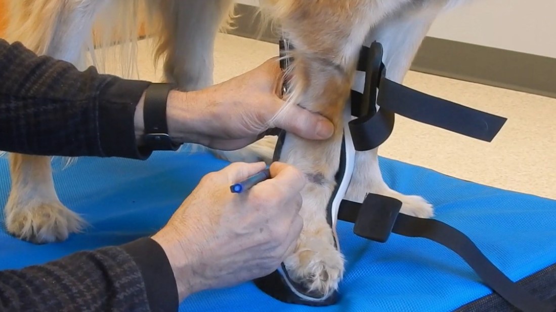
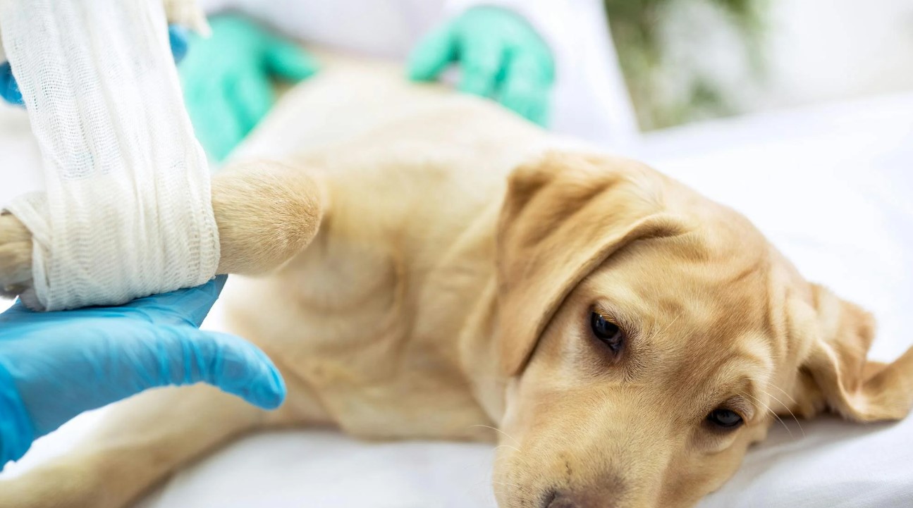
- Postoperative Care: After surgery, the dog’s leg is placed in a plaster cast to prevent any movement. The cast is typically left in place for 1.5 to 2 months, during which time the bone fusion is monitored with X-rays.
Postoperative Recovery and Rehabilitation
Recovery from arthrodesis is a long process that requires careful management to ensure successful bone fusion. After surgery, the dog must wear a cast or splint to keep the affected limb immobilized. Physical activity is restricted, and the dog must be closely monitored to prevent any strain on the joint while it heals.
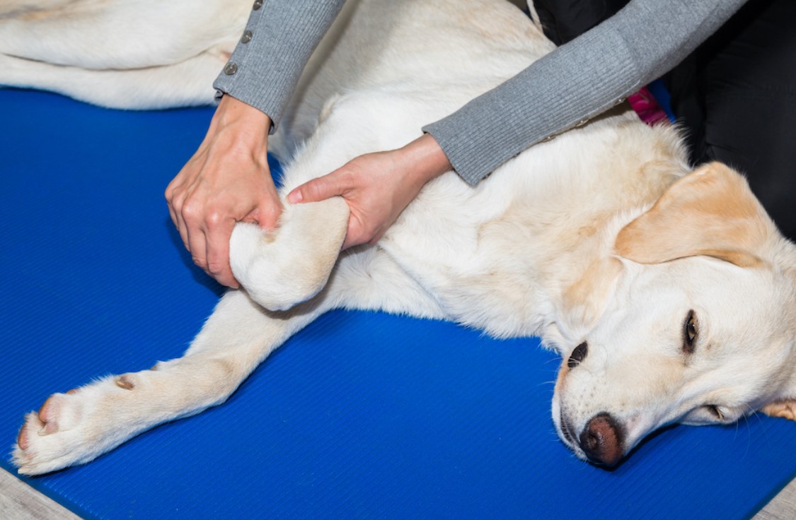
X-rays are taken at regular intervals to monitor the progress of bone fusion. Once the veterinarian confirms that the bones have successfully fused, the cast or splint can be removed. Rehabilitation exercises, such as controlled walking and swimming, may be recommended to help the dog regain strength in the affected limb.
Owners should be aware that while the joint will no longer move, most dogs adapt well to the loss of mobility and can live pain-free and functional lives after arthrodesis.
Advantages and Disadvantages of Arthrodesis
The primary benefits of arthrodesis include:
– Pain Relief: Arthrodesis eliminates the painful movement of bones within the joint, providing significant relief for dogs suffering from chronic joint pain.
– Restoration of Limb Function: By stabilizing the bones in a functional position, arthrodesis can restore partial function to the affected limb, allowing the dog to use the limb more effectively.
However, there are also important considerations:
– Loss of Joint Mobility: The fused joint will no longer be able to move, which may limit the dog’s range of motion. While most dogs adapt to this change, owners should be aware that the animal will not regain full limb function.
– Lengthy Recovery: The recovery process after arthrodesis is long and requires careful monitoring. Dogs must be kept immobilized for several months to ensure proper healing, and owners must be committed to managing the dog’s rehabilitation.
Arthrodesis in Dubai: Veterinary Expertise and Clinics
For dog owners in Dubai seeking arthrodesis surgery, it is essential to consult experienced veterinarians who specialize in orthopedic care. Clinics such as Vets in the City are well-known for their expertise in diagnosing and treating complex musculoskeletal issues in dogs.
At Vets in the City, a team of board-certified orthopedic surgeons provides advanced diagnostic techniques, surgical interventions, and comprehensive postoperative care. The clinic offers a full range of services, from initial consultation to surgery and rehabilitation, ensuring that every pet receives the highest standard of care. Whether your dog is suffering from a traumatic injury, degenerative joint disease, or other orthopedic issues, Vets in the City is equipped to provide personalized treatment plans tailored to each pet’s unique needs.
Conclusion
Arthrodesis is a critical surgical option for dogs suffering from severe joint damage or chronic pain. While it results in the permanent immobilization of the affected joint, the procedure offers significant pain relief and restores partial functionality to the limb. Proper postoperative care, rehabilitation, and close monitoring are essential to ensuring successful outcomes. For pet owners in Dubai, seeking out experienced orthopedic veterinarians, such as those at Vets in the City, can help ensure the best possible results for dogs requiring this procedure.

Football fan, shiba-inu lover, DJ, hand letterer and identity designer. Operating at the fulcrum of modernism and elegance to craft experiences that go beyond design. Let’s chat.